
Data description
Unstructured noise
Unstructured noise in the process of microscopy image acquisition refers to random variations in the pixel values of an image that do not follow a predictable pattern or structure. This type of noise can arise from various sources.
Most dominant sources of noise (such as Poisson shot noise and additive Gaussian read-out noise) are usually assumed to fall into this category.
C. Broaddus, A. Krull, M. Weigert, U. Schmidt, and G. Myers, "Removing
Structured Noise with Self-Supervised Blind-Spot Networks," 2020
Datasets for unstructured noise leaderboards
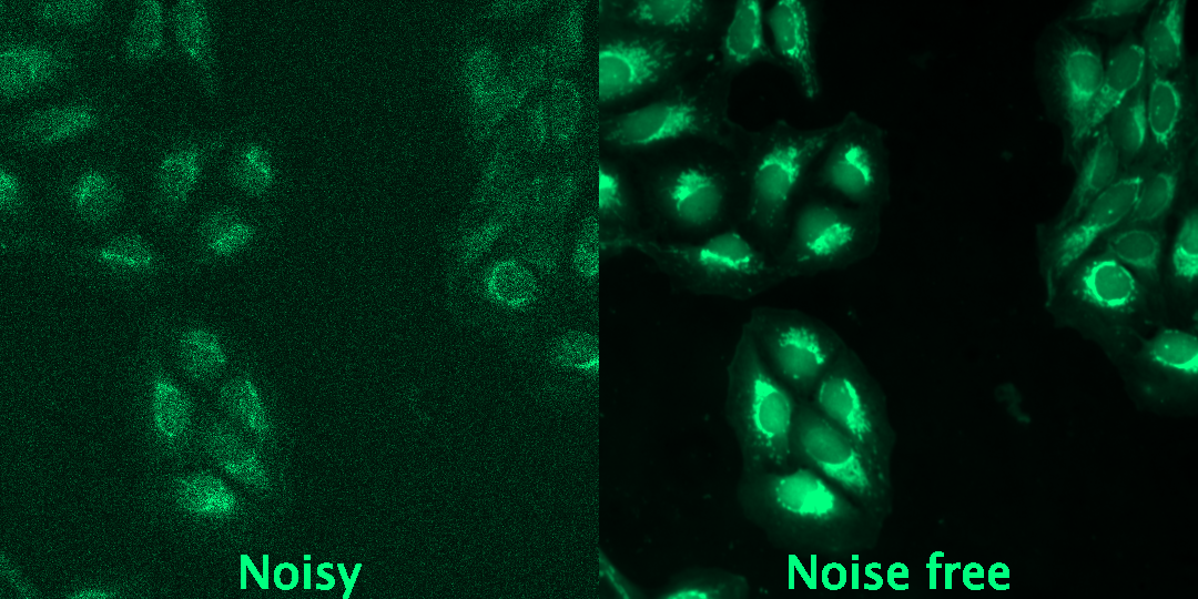
Unstructured noise 1: JUMP Cell painting Datasets
The JUMP Cell Painting image datasets are large, public reference data from chemical and genetic perturbations. It contains a comprehensive collection of cellular imaging data aimed at advancing drug discovery through high-dimensional data analytics and image analysis. Cell Painting is a high-content, fluorescence imaging technique that uses a series of dyes to stain different components of the cell, such as the nucleus, cytoplasm, mitochondria, Golgi apparatus, endoplasmic reticulum, and actin cytoskeleton.
In this challenge, we provide a subset of images from the JUMP Cell painting dataset subjected to synthetic noise. We apply the Gaussian noise and Poisson to simulate the detector noise, which arises due to the electronics used to detect light, e.g., cameras or photodetectors.
You can find the training dataset here: https://zenodo.org/records/10912386
We used the JUMP Cell Painting datasets (Chandrasekaran et al., 2023), available from the Cell Painting Gallery on the Registry of Open Data on AWS (https://registry.opendata.aws/cellpainting-gallery/).
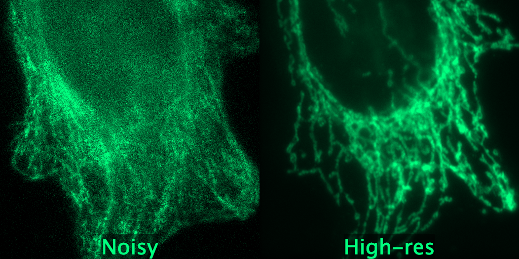
Unstructured noise 2: W2S: Microscopy Data with
Joint Denoising and Super-Resolution for Widefield to SIM Mapping
The W2S dataset is acquired using conventional fluorescence widefield and SIM imaging. A set is composed of noisy low-resolution (LR) widefield images with different noise levels, a noise-free LR image, and a corresponding high-quality HR SIM image.
You can find the training dataset here: https://zenodo.org/records/10925783
Zhou, R., El Helou, M., Sage, D., Laroche, T., Seitz, A., Süsstrunk, S. (2020). W2S: Microscopy Data with Joint Denoising and Super-Resolution for Widefield to SIM Mapping. In: Bartoli, A., Fusiello, A. (eds) Computer Vision – ECCV 2020 Workshops. ECCV 2020. Lecture Notes in Computer Science(), vol 12535. Springer, Cham. https://doi.org/10.1007/978-3-030-66415-2_31
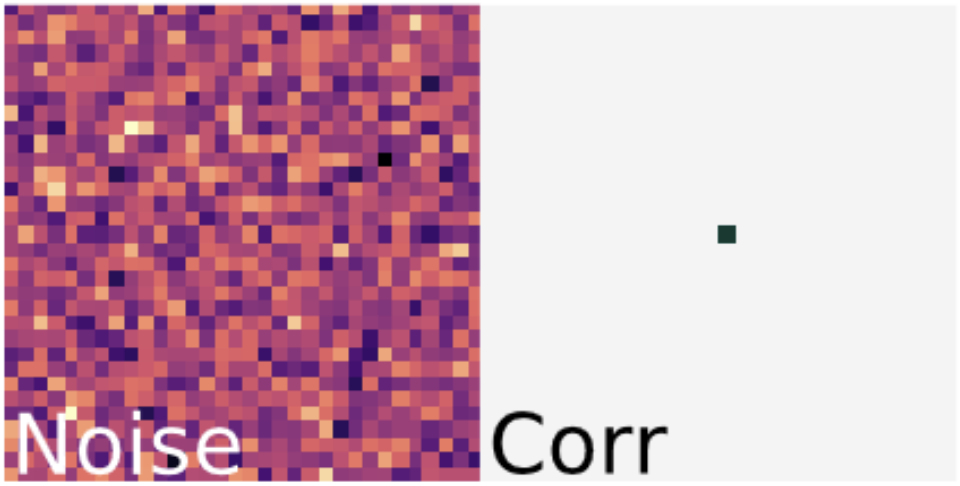
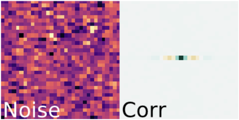
Structured noise
Structured noise in the context of microscopy image acquisition refers to patterns of interference or artifacts that are not random but have a specific structure or organization. This type of noise can arise from various sources within the imaging process, such as imperfections in the optical system and sensor noise.
This type of noise is highly correlated between neighbouring pixels, creating regular patterns in the image.
C. Broaddus, A. Krull, M. Weigert, U. Schmidt, and G. Myers, "Removing Structured Noise with Self-Supervised Blind-Spot Networks," 2020
Datasets for structured noise leaderboards
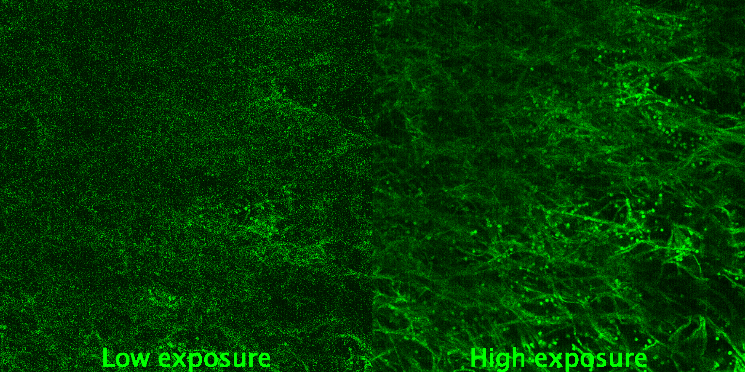
Structured noise 1: Fluorescence
Microscopy Datasets for Training Deep Neural Networks by Hagen et al.
The authors provide a high-quality dataset comprising whole images under low-SNR and high-SNR exposure settings. It covers pairs of images acquired with different exposure times (or, in the case of confocal microscopy, different laser power and detector gain settings).
We provide a subset of low-snr images subject to structured noise for training.
You can find the training dataset here:
https://zenodo.org/records/10925855

Guy M Hagen, Justin Bendesky, Rosa Machado, Tram-Anh Nguyen, Tanmay Kumar, Jonathan Ventura, Fluorescence microscopy datasets for training deep neural networks, GigaScience, Volume 10, Issue 5, May 2021, giab032, https://doi.org/10.1093/gigascience/giab032
Structured noise 2: SUPPORT (Statistically unbiased prediction enables accurate
denoising of voltage imaging data) method
This method provides a volumetric dataset that contains Penicillium imaged with confocal Microscopy. It was imaged using two different recording settings to generate a pair of low-SNR and high-SNR volumes.
We provide a stack of images with low-snr subject to structured noise for training.
You can find the training dataset here: https://zenodo.org/records/10925939
Eom, M., Han, S., Park, P. et al. Statistically unbiased prediction enables accurate denoising of voltage imaging data. Nat Methods 20, 1581–1592 (2023). https://doi.org/10.1038/s41592-023-02005-8